Peptide Receptor Binding: How Peptides Interact with Their Targets
A primer on the molecular pharmacology of peptide-receptor binding, covering receptor types, binding kinetics, agonism vs. antagonism, and what binding affinity data means for interpreting research.
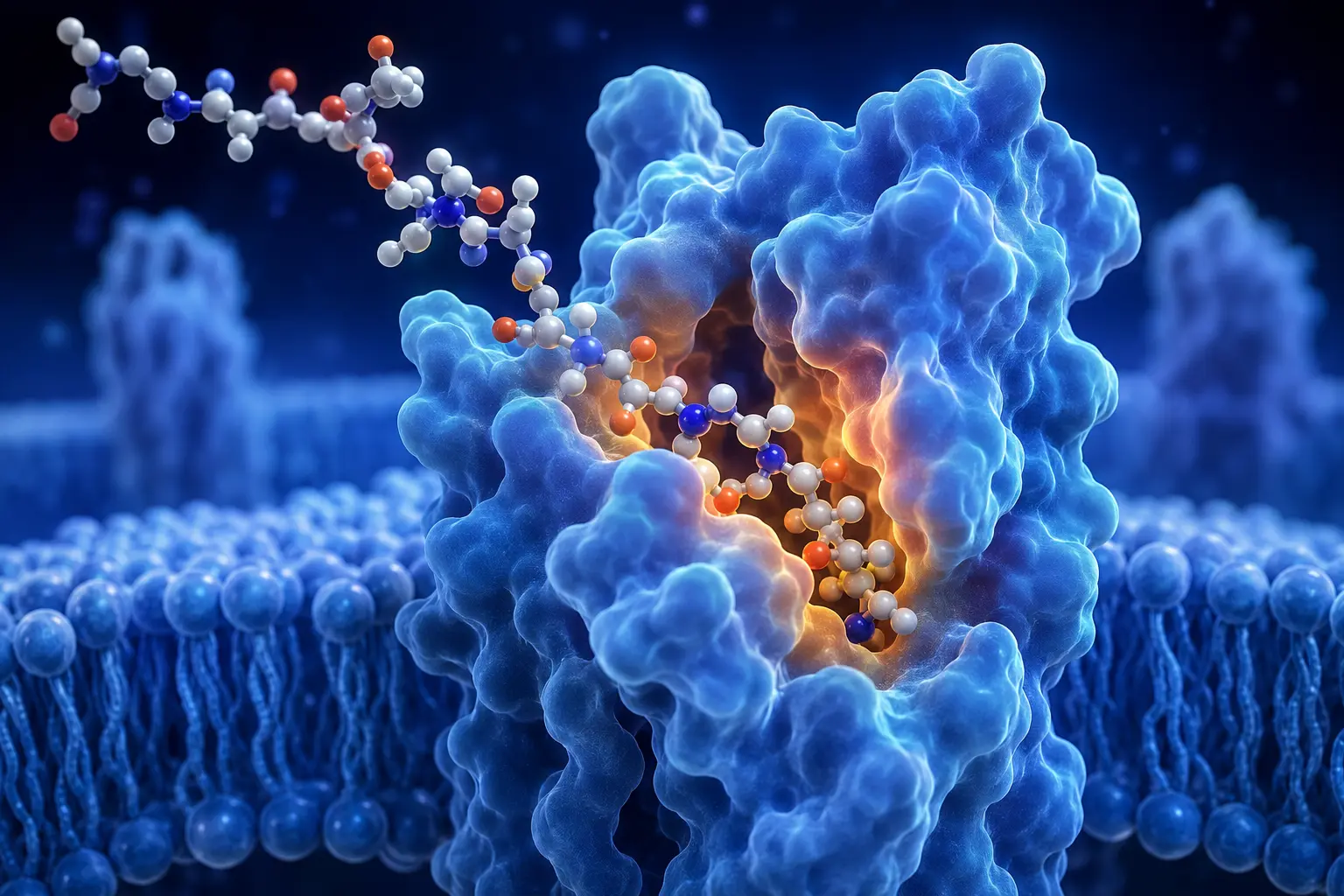
Peptide activity in biological systems depends fundamentally on the interaction between the peptide and its molecular target — most commonly a cell-surface receptor. Understanding receptor binding principles is essential for interpreting peptide pharmacology research, evaluating binding affinity data, and understanding why structurally similar peptides can have dramatically different effects.
Receptor Types for Peptide Ligands
The vast majority of peptide hormones and signaling molecules act through G protein-coupled receptors (GPCRs). GPCRs are seven-transmembrane domain proteins that, when activated by a ligand, couple to intracellular G proteins (Gs, Gi, Gq, and others), initiating signal transduction cascades.
Why predominantly GPCRs? The GPCR superfamily represents the largest and most versatile receptor family in mammals, with over 800 members. Their structure allows diverse ligand binding sites (some peptides bind the extracellular domain, others insert into the transmembrane bundle), and their downstream signaling is highly adaptable.
Some peptide ligands also act through:
- Receptor tyrosine kinases (RTKs): Insulin and IGF-1 receptors are prominent examples
- Cytokine receptors: Erythropoietin, growth hormone, and prolactin receptors
- Enzyme-linked receptors: Natriuretic peptide receptors (guanylyl cyclase type)
- Nuclear receptors: Some lipid-soluble bioactive peptide-derived molecules
Binding Affinity: Kd and Ki
Binding affinity quantifies how tightly a ligand binds to a receptor. Two commonly reported parameters:
Kd (dissociation constant): The concentration of ligand at which 50% of receptors are occupied at equilibrium. Lower Kd = higher affinity. A peptide with Kd = 1 nM binds its receptor 1,000-fold more tightly than one with Kd = 1 μM.
Ki (inhibition constant from competition binding assays): Derived from competitive radioligand binding experiments, where the test compound displaces a radiolabeled reference ligand. Ki has the same units and interpretation as Kd.
These values are measured in vitro using isolated receptor preparations (membranes, cell lysates, or expressed receptors in cell lines). They describe thermodynamic binding properties, not biological activity.
Critical limitation: High binding affinity does not guarantee biological effect. A high-affinity antagonist binds tightly but produces no signal. A compound can have high affinity but poor selectivity (binding many receptors). In vivo bioavailability and tissue distribution further separate binding affinity from observed biological activity.
Agonism, Antagonism, and Partial Agonism
When a peptide binds a receptor, it can produce different functional outcomes:
Full agonist: Binds and activates the receptor to the same maximum response as the endogenous ligand. GLP-1 receptor agonists in current pharmaceutical use are typically full agonists designed to produce maximal receptor activation.
Partial agonist: Binds and activates the receptor but produces a submaximal response even at receptor saturation. Can act as an antagonist in the presence of a full agonist (occupies receptors without producing full signal). Some research peptide analogs are partial agonists.
Antagonist: Binds without activating. Competitively blocks the endogenous ligand or other agonists. Clinically useful antagonists include GnRH antagonists (cetrorelix, ganirelix) used in IVF protocols.
Inverse agonist: Binds and reduces receptor activity below its constitutive (basal) level. Less common but relevant for receptors with significant constitutive activity.
Receptor Selectivity
When a peptide is described as "selective" for a receptor, this means it has a significantly higher affinity for the target receptor than for other related receptors. Selectivity is always relative and context-dependent.
For research purposes, selectivity is important because it helps researchers attribute observed biological effects to specific receptor mechanisms. For further reading on how specific growth hormone secretagogues leverage receptor selectivity in research design, see our overview of the GHS class.
Selectivity data is typically presented as a ratio of Ki values:
Selectivity ratio = Ki(off-target receptor) / Ki(target receptor)
A selectivity ratio of 100 means the compound is 100-fold more potent at the target than the off-target receptor. Whether this is sufficient selectivity for clean research use depends on the relative expression levels of both receptors in the tissue studied.
Downstream Signaling: From Binding to Effect
Receptor binding initiates intracellular signaling cascades. For a Gs-coupled receptor (like GHS-R1a for GH release):
- Ligand binds receptor
- Receptor changes conformation, activating Gαs subunit
- Gαs activates adenylyl cyclase
- cAMP levels rise
- cAMP activates protein kinase A (PKA)
- PKA phosphorylates target proteins (including those involved in GH granule exocytosis)
At each step, there is amplification (one receptor activation can generate many cAMP molecules), regulation (phosphodiesterases degrade cAMP), and cross-talk with other signaling pathways. This means the relationship between receptor occupancy and biological effect is not linear.
Receptor Desensitization
Prolonged or repeated receptor activation typically leads to desensitization — a reduced response to the same stimulus over time. Mechanisms include:
Phosphorylation: Activated GPCRs are phosphorylated by GRKs (GPCR kinases), reducing coupling efficiency to G proteins.
Beta-arrestin recruitment: Phosphorylated receptors recruit beta-arrestin, which both uncouples the receptor from G proteins and initiates receptor internalization.
Receptor internalization and downregulation: Receptors are removed from the cell surface via endocytosis. After internalization, receptors can be recycled (resensitization) or degraded (downregulation of receptor number).
This desensitization biology is directly relevant to research peptide use. The tachyphylaxis (diminishing response) observed with hexarelin at high doses reflects receptor desensitization. Understanding this phenomenon prevents misinterpreting reduced responses as loss of compound activity.