Follistatin: TGF-β Superfamily Antagonist in Research
Follistatin research: a TGF-β antagonist that inhibits myostatin, covering the distinction between the follistatin protein and synthetic peptide fragments.
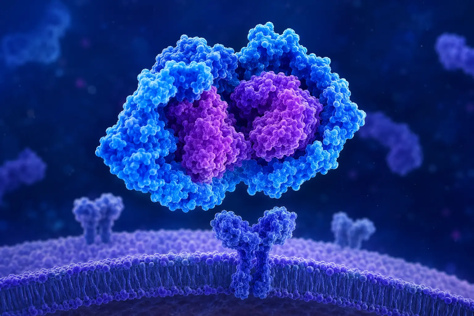
Follistatin is a naturally occurring glycoprotein — not a short synthetic peptide — that functions as an extracellular binding protein for several members of the TGF-β (transforming growth factor-beta) superfamily, most notably activin and myostatin. It is included in peptide research discussions primarily because of its role in myostatin inhibition and because synthetic peptide fragments derived from its active regions are sometimes used as research compounds.
Researchers reading about follistatin in research contexts need to distinguish carefully between:
- Recombinant human follistatin protein (rhFST) — a large glycoprotein
- Follistatin gene therapy approaches (delivery of FST gene via viral vectors)
- Short synthetic peptide fragments purported to mimic follistatin binding activity
These are entirely different compounds with different evidence bases.
Follistatin Biology
Follistatin (FST) is encoded by the FST gene and circulates in plasma as a 35–40 kDa glycoprotein (molecular weight varies with glycosylation). The major isoforms are FST288 (binds heparan sulfate proteoglycans, largely cell-bound) and FST315 (circulates in plasma).
Follistatin binds its ligands — primarily activin A, activin B, and GDF-8 (myostatin) — with high affinity and prevents them from binding their functional receptors. It does not compete at the receptor level; instead, it neutralizes the ligands before receptor contact.
Myostatin: The Target of Interest for Muscle Research
Myostatin (GDF-8) is a potent inhibitor of skeletal muscle growth. Its role was dramatically illustrated in myostatin knockout mice, which develop approximately twice the muscle mass of normal mice through both increased muscle fiber number (hyperplasia) and fiber size (hypertrophy). Similar phenotypes have been observed in naturally occurring myostatin-null cattle (Belgian Blue, Piedmontese breeds) and rarely in humans with myostatin mutations.
Because follistatin can bind and neutralize myostatin, the hypothesis that increasing follistatin may promote muscle growth has attracted substantial research interest. Animal studies using follistatin gene therapy in mice and non-human primates have reported impressive increases in muscle mass.
What Animal Research Has Examined
Follistatin gene therapy in mice: Studies overexpressing follistatin in mouse muscle have reported substantial increases in muscle mass. Some studies have shown that this muscle mass increase may persist even in disease models.
Follistatin + exercise models: Some research has examined whether follistatin may amplify resistance exercise-induced muscle hypertrophy in animal models.
Age-related muscle loss (sarcopenia) models: Animal research has examined whether follistatin supplementation might attenuate age-related muscle loss, based on the observation that myostatin/activin signaling is relatively increased in aged muscle.
Human Research
Human data on follistatin intervention is extremely limited. The primary human data comes from:
Follistatin gene therapy clinical trials: A small open-label trial in Becker muscular dystrophy patients examined intramuscular injection of an AAV vector delivering FST344 (a modified follistatin isoform). The Mendell group at Nationwide Children's published results showing some safety and preliminary efficacy signal in this rare disease context. This is not generalizable to healthy individuals.
Measurement studies: Research examining how exercise, diet, or other interventions affect endogenous follistatin levels in humans. These are observational data about the natural follistatin system, not supplementation studies.
No well-designed RCT examining exogenous follistatin protein administration in humans for any indication has been published as of this writing.
Synthetic Follistatin Peptide Fragments
Short synthetic peptides marketed as "follistatin peptides" are not follistatin. They are fragments of the follistatin sequence — most commonly identified as coming from particular functional regions of the protein.
Critical points for researchers:
- A peptide fragment does not automatically replicate the activity of the parent protein
- The binding surfaces on follistatin involved in myostatin neutralization are discontinuous structural epitopes — they depend on the three-dimensional folded structure of the protein A short linear peptide fragment cannot necessarily recapitulate the binding geometry of the folded protein.
- Published evidence specifically characterizing synthetic follistatin fragment activity in robust biological systems is extremely limited
Claims that short synthetic "follistatin peptides" produce myostatin inhibition comparable to follistatin protein should be evaluated with this structural biology context in mind.
Regulatory and Safety Considerations
Myostatin pathway manipulation — through any mechanism — raises potential safety considerations beyond muscle growth, including effects on cardiac muscle, adipose tissue metabolism, bone, and reproductive biology (follistatin regulates FSH through activin B antagonism, which has gonadal implications). These potential off-target effects have not been characterized for long-term systemic intervention in humans.
References
- 1.Mendell JR, Sahenk Z, Al-Zaidy S, Rodino-Klapac LR, Kissel JT, Prior TW, Burke RT, Blaschak K, D'Ambrosio P, Kaspar BK. “Follistatin Gene Therapy for Sporadic Inclusion Body Myositis Improves Functional Outcomes.” Molecular Therapy. 2017;25(4):870-879. doi:10.1016/j.ymthe.2017.02.015 [PubMed]