IGF-1: Insulin-Like Growth Factor in Research Context
An overview of IGF-1 (insulin-like growth factor 1), its role as the primary downstream mediator of growth hormone action, and what research examines regarding its effects on tissues and metabolism.
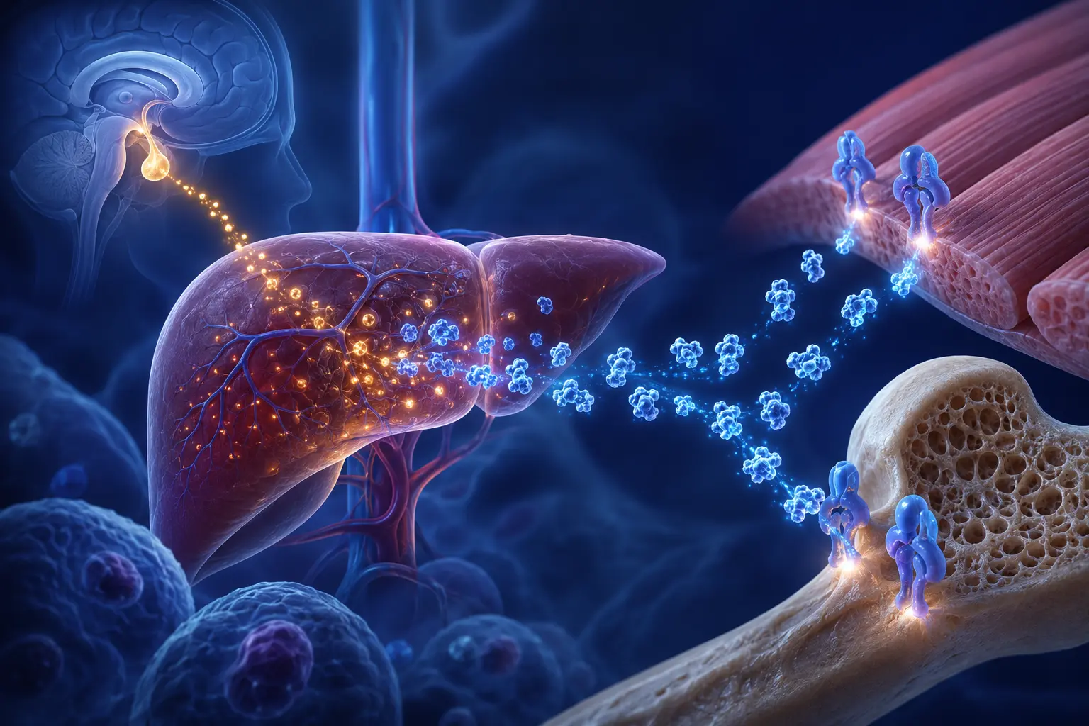
IGF-1 (Insulin-Like Growth Factor 1) is a 70-amino-acid single-chain polypeptide produced primarily by the liver in response to growth hormone (GH) signaling. It is one of the primary mediators through which GH exerts its anabolic effects on peripheral tissues. Understanding IGF-1 biology is essential for interpreting research on growth hormone secretagogues, as IGF-1 is a commonly measured endpoint in that literature.
The GH-IGF-1 Axis
Growth hormone is released from the pituitary gland in a pulsatile pattern, with the largest pulses typically occurring during deep sleep. GH acts on receptors in the liver (and other tissues) via the JAK2/STAT5b signaling pathway to stimulate IGF-1 synthesis and secretion.
Circulating IGF-1 then acts on IGF-1R (the IGF-1 receptor), which belongs to the same receptor family as the insulin receptor. IGF-1R activation initiates signaling cascades including PI3K/Akt and MAPK/ERK pathways, which regulate cell growth, proliferation, survival, and metabolism.
IGF-1 also feeds back to suppress GH release from the pituitary (negative feedback), forming the GH-IGF-1 axis.
IGF-1 Binding Proteins
A critical feature of IGF-1 that researchers must account for is that the majority of circulating IGF-1 (over 95%) is bound to one of six IGF-binding proteins (IGFBPs 1–6). These binding proteins:
- Extend IGF-1's plasma half-life from minutes to hours
- Serve as transport proteins and tissue-specific reservoirs
- Can inhibit or enhance IGF-1 activity depending on context
- Are independently regulated by various hormones and nutritional signals
When research measures "total IGF-1" in plasma, it is measuring the sum of bound and free IGF-1. Only free IGF-1 can bind IGF-1R. The ratio of free to bound IGF-1 is influenced by IGFBP levels, which in turn are regulated by many factors including insulin levels, nutritional status, and exercise. This complexity is often not fully addressed in peptide research studies.
What Studies Have Examined Regarding IGF-1 Effects
The biological activities under study with IGF-1 are extensive. Researchers have examined potential effects on:
Skeletal muscle: Cell culture and animal studies have examined whether IGF-1 may promote myocyte (muscle cell) hypertrophy and potentially inhibit atrophy. IGF-1 has been studied in models of muscle wasting including sarcopenia, cachexia, and disuse atrophy.
Bone: IGF-1 receptors are expressed on osteoblasts (bone-forming cells). Research has examined whether IGF-1 signaling may influence bone density and geometry. GH-deficient individuals often have reduced bone density, and GH/IGF-1 replacement has been studied in this context.
Glucose metabolism: IGF-1 can activate insulin receptors at high concentrations due to structural similarity to insulin. Research has examined insulin-sensitizing effects of IGF-1, though its overall role in glucose metabolism is complex and context-dependent.
Neurological applications: IGF-1 crosses the blood-brain barrier (unlike GH) and IGF-1R is widely expressed in the CNS. Research has examined potential neuroprotective effects in various models, and IGF-1 is being studied in clinical trials for ALS and some other neurological conditions.
IGF-1 and Cancer Risk: An Important Consideration
The growth-promoting properties of IGF-1 — the same properties that make it interesting for anabolic research — raise concern about oncogenic potential. IGF-1R signaling is a recognized promoter of cell proliferation and survival, and elevated IGF-1 levels have been associated with increased risk for several cancers in epidemiological studies, particularly colorectal cancer and premenopausal breast cancer.
The IGF-1/cancer relationship is dose- and context-dependent, and the epidemiological associations do not establish causation. However, researchers and clinicians are obligated to consider this relationship when evaluating long-term IGF-1 elevation, whether from GH replacement, GH secretagogue use, or exogenous IGF-1 administration.
Researchers investigating GH secretagogues often measure IGF-1 as an indicator of GH action, but reporting IGF-1 elevations without acknowledging the oncogenic context is an incomplete presentation of the evidence.
Normal Reference Ranges and Age Decline
IGF-1 levels follow a characteristic life-course pattern:
- Low in early childhood
- Peak during puberty (adolescent growth spurt)
- Gradual decline through adulthood (approximately 14% per decade after age 30)
- Significantly reduced in late life
This age-related decline in IGF-1 has been interpreted by some researchers as a potential contributor to age-related loss of muscle mass and bone density. Others have argued that lower IGF-1 in older age may actually be protective against cancer risk. This debate is not resolved in the literature.
Reference ranges vary by laboratory and assay, but typical adult ranges are approximately 100–300 ng/mL, with substantial age and sex variation.
References
- 1.Le Roith D, Bondy C, Yakar S, Liu JL, Butler A. “The somatomedin hypothesis: 2001.” Endocrine Reviews. 2001;22(1):53-74. doi:10.1210/edrv.22.1.0419 [PubMed]
- 2.Chan JM, Stampfer MJ, Giovannucci E, Gann PH, Ma J, Wilkinson P, Hennekens CH, Pollak M. “Plasma insulin-like growth factor-I and prostate cancer risk: a prospective study.” Science. 1998;279(5350):563-566. doi:10.1126/science.279.5350.563 [PubMed]
- 3.Hankinson SE, Willett WC, Colditz GA, Hunter DJ, Michaud DS, Deroo B, Rosner B, Speizer FE, Pollak M. “Circulating concentrations of insulin-like growth factor I and risk of breast cancer.” Lancet. 1998;351(9113):1393-1396. doi:10.1016/S0140-6736(98)01346-2 [PubMed]